Case Presentation
A 32 year female, with no co morbidities, case of primary infertility due to partial vaginal agenesis/ bicornuate uterus, H/o traumatic hematometra drainage followed by which the patient was
having delayed cycles, and fecal discharge from vagina.
Patient was initially treated in A4 Fertility centre, Chennai in April 2018
RVF repair + diversion transverse loop colostomy done on April 2018
5/9/22
C/o amenorrhea for 2 years
TVS of pelvis done on 5/9/22 showed normal uterus, endometrial cavity, right ovary, adnexae, pouch of douglas
Per vaginal examination:
RVF Rent present, connected to rectum
13/10/22
Upon review in MGMCRI general physical examination was unremarkable
Per rectal examination:
Rectovaginal fistula present at 12'o clock position 1cm from anal verge around ~4-5cm in length
Rectum empty
No active bleeding/ discharge
CECT Abdomen:
Recto-uterine fistula as described Left kidney is malrotated and fused to lower pole of the right kidney -Cross fused malrotated kidney hypoattenuating lesion 4x3mm with CT attenuation value 24HU in right lobe of liver (Segment VI). Lesion shows intense enhancement in arterial phase and retention of contrast in venous phase.
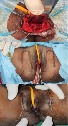
Patient underwent dismantling of rectovaginal fistula with primary repair of rectovaginal fistula, interposition bilateral pudendal artery perforator flapon 15/10/22
Post operative period uneventful.
Planned for reversal of colostomy
Discussion
It is one of the most difficult surgical problems in colorectal surgery.
Previous failed attempts will cause difficulty in next further attempts
Many factors must be considered when choosing an operative approach to rectovaginal fistulae. The number and type of previous repairs, patient's risk factors, additional concomitant fistulae, and sphincter integrity are all concerns. Of these factors, the cause of the rectovaginal fistula and the status of the external anal sphincter are the most important.
Gracilis Flap
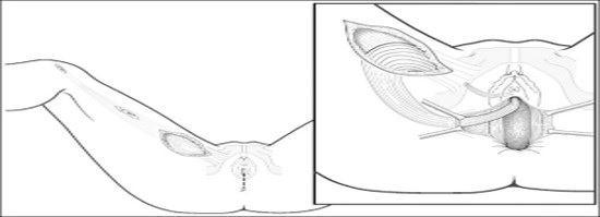
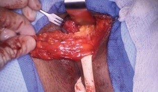
The gracilis muscle is often used as an interposition flap. The procedure can be performed by the fistula surgeon or in conjunction with a plastic surgeon with similar results.The surgery is typically performed in the lithotomy position, with the gracilis muscle being harvested from a 8 to 10 cm incision on the medial thigh. A perineal incision is then made, dissecting well above the fistula itself with care taken not to violate the rectal or vaginal mucosa. The pedicled muscle flap is tunneled to the perineal incision and secured into place with absorbable sutures.
Paint
Reported primary healing rates for gracilis interposition grafts generally range from 75 to 92%.It should be noted that most case series on this topic involve complex fistulae with multiple previous attempts at repair. A large number of these fistulae were secondary to Crohn disease and radiation injury, with a small number being secondary to obstetrical injury. In addition, most case series employed routine fecal diversion.
Martius flap
Another well-described tissue transfer and local flap repair is the use of the Martius or bulbocavernosus flap. Using a longitudinal incision over the labia majora, skin flaps are raised laterally and medially, with dissection continuing to the periosteum of the pubis and to the pubic symphysis. Once the entire fat pad with the bulbocavernosus muscle is mobilized, the anterior aspect is cut and used as a vascularized pedicled flap (perineal branch of the pudendal artery) and tunneledsubvaginally. The flap is then sutured to the posteriorvaginal wall to interpose it over the closed rectal aspect of the fistula.
Aartsen and Sindram [5] initially reported 100% success in 14 patients with fistulae secondary to radiation damage, but after a 10-year follow-up, 8 of the 14 patients required diversion for continued damage from radiation. Pitel et al [5] reported a 65% overall healing rate for Martius flap, with a 50% rate in patients with Crohn disease.
Bilateral Internal pudendal artery perforator flap (IPAP flap)
After complete debridement of the fistula and the surrounding unhealthy tissue, and mobilizing rectum the exact localization of the flap to be elevated was determined via tracing of the IPAP The flaps were partitioned into 2 triangles, one on each side. The area of triangles were determined considering the size of the rectal opening and the distance between the vulva and the vaginal opening. The proximal part of the triangle was de-epithelialized and folded with the subcutaneous fat sides facing each other, and was then transposed between the vagina and rectum to Replace the damaged rectovaginal septum. As a result, both the vaginal and rectal openings of the fistula were covered with a well-vascularized skin paddle
The utilization of IPAP flap for recurrent RVF is a reliable, safe, easy-to-execute option, which has the same urogenital dermatomal supply and low donor-site morbidity. Its versatile nature allows reconstruction of concomitant perineal skin defect with minimal anatomical disturbance and hence should be considered as a valuable tool in the armamentarium of RVF management.
References
- Goldaber KG, Wendel PJ, McIntire DD, Wendel GD. Postpartum perineal morbidity after fourth-degree perineal repair. Am J Obstet Gynecol. 1993; 168: 489-493.
- Homsi R, Daikoku NH, Littlejohn J, Wheeless CR. Episiotomy: risks of dehiscence and rectovaginal fistula. Obstet Gynecol Surv. 1994; 49: 803-808.
- Wall LL, Karshima JA, Kirschner C, Arrowsmith SD. The obstetric vesicovaginal fistula: characteristics of 899 patients from Jos, Nigeria. Am J Obstet Gynecol. 2004; 190: 1011-1019.
- Pitel S, Lefevre JH, Parc Y, Chafai N, Shields C, Tiret E. Martius advancement flap for low rectovaginal fistula: short- and long-term results. Colorectal Dis. 2011; 13: e112-e115.
- Aartsen EJ, Sindram IS. Repair of the radiation induced rectovaginal fistulas without or with interposition of the bulbocavernosus muscle (Martius procedure). Eur J Surg Oncol. 1988; 14: 171-177.