Introduction
Brachymetatarsia is a congenital deformity characterized by the shortening of one or more metatarsal bones, often the fourth metatarsal [1,2]. Due to misalignment and overload on the surrounding joints and tissues, this condition not only alters the appearance of the foot but can also cause pain and affect the biomechanical function of the foot [1]. Surgical treatment is considered the most effective option to correct the deformity and restore normal foot functionality, despite several conservative treatments being described [2,3].
The primary goal of surgical treatment for brachymetatarsia is to restore the length and alignment of the affected metatarsal, improving both the biomechanical function and aesthetic appearance of the foot [3]. Two main surgical techniques are described in the literature: one-stage osteotomy (shortening through immediate distraction with bone graft) and gradual lengthening using external distraction devices [1,4].
Gradual lengthening with external distraction, also known as callotasis or the Ilizarov technique, has gained popularity in recent decades due to its ability to correct more severe cases of brachymetatarsia and reduce complications associated with bone grafts [3,5]. This procedure involves placing an external fixation device on the affected metatarsal, followed by an osteotomy. The bone is then gradually lengthened through controlled distraction of the device, typically at a rate of 0.5-1 mm per day [6-8].
One-stage osteotomy, also known as acute metatarsal lengthening, involves cutting the affected metatarsal bone and inserting an intercalary bone graft, usually taken from the iliac bone or using bone substitutes [2,5]. Among the one-stage lengthening techniques, the following approaches are described:
Wedge osteotomy with intercalary bone graft: This involves performing a wedge osteotomy on the affected metatarsal, and the resulting defect is filled with a bone graft (autologous, allogeneic, or banked). Internal fixation with plates and screws is usually used. This technique is indicated for lengthenings up to 15 mm. Complications include an increased risk of nonunion, bone graft failure, or delayed consolidation [9].
Segmental osteotomy with internal fixation: This technique is based on a segmental osteotomy of the metatarsal with internal fixation, avoiding the use of bone grafts. It is an acute lengthening procedure where the desired length is achieved in a single surgical stage using fixation with plates, screws, or intramedullary nails. It is indicated for lengthenings of less than 10 mm. This technique carries a higher risk of stress at the osteotomy site, delayed consolidation, and misalignment [10].
L-shaped osteotomy: In this technique, an “L” shaped osteotomy is performed, which facilitates both mechanical stability of the bone and one-stage correction. Internal fixation with screws or a special plate is used. It is suitable for small to moderate lengthenings, with a lower risk of mechanical failure. However, it presents difficulties in achieving lengthenings greater than 15 mm and possible overload on the metatarsophalangeal joints [11].
Z-plasty metatarsal osteotomy: A “Z” shaped osteotomy is performed on the metatarsal to facilitate longitudinal expansion. This approach allows for a more precise correction of length and also facilitates realignment in the case of angular deformity. It is useful for moderate lengthenings and is an option for patients with mild to moderate angular deformities. Complications include issues related to fixation and postoperative alignment [12].
Oblique osteotomy with banked bone graft: This technique involves an oblique osteotomy of the affected metatarsal, followed by the interposition of a banked bone graft to achieve lengthening. Internal fixation with plates or screws is used to stabilize the graft and the recipient bone. It is suitable for lengthenings greater than 15 mm, where graft stability is essential for the success of the procedure. It carries a high risk of bone necrosis and graft failure [13].
The objective of this article is to present a case of bilateral brachymetatarsia treated using the oblique technique with an autologous calcaneal graft. In one of the cases, digital necrosis occurred due to excessive lengthening. Ultimately, the results obtained were satisfactory.
Materials and methods
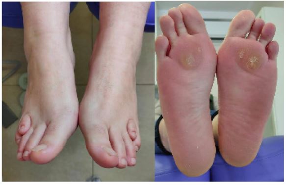
A 41-year-old woman presented to the podiatry clinic on April 8, 2022, due to pain in the plantar area of the third metatarsal. She reported no allergies or significant medical history, although she mentioned similar deformities in her family. Her risk assessment score according to the Autar scale was 12 points, she was overweight, and her SF-36 score was 84 points. Neurological and vascular examinations were normal.
Clinically, first ray insufficiency was observed, leading to mild hallux abductus. Additionally, overload was identified in the second and third metatarsals, secondary to bilateral brachymetatarsia of the fourth metatarsal. Biomechanical evaluation revealed a valgus flatfoot (FPI+7) with first ray insufficiency and overload on the central rays.
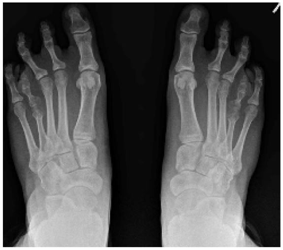
Radiological studies revealed a slight deviation of the first metatarsophalangeal segment, with an increase in intermetatarsal angles (14O on the left foot and 12O on the right), interphalangeal angles (16O on the left foot and 14O on the right), and hallux abductus valgus angles (24O on the left foot and 20O on the right). No alterations were found in the PASA or DASA angles. The metatarsal parabola angle, according to Merchan, measured 137O on the left foot and 134O on the right.
Regarding the brachymetatarsia, the fourth metatarsal was thin, with slender cortices and in dorsiflexion, showing a shortening of 8 mm on the left foot and 6 mm on the right. The M4-M5 intermetatarsal angle was 9O on the left foot and 8O on the right.
On June 4, 2022, surgery was performed on the left foot. An Austin Youngswick osteotomy with slight plantarflexion and a 3 mm dorsal wedge was performed, along with a cylindrical Akin osteotomy removing a 4 mm wedge. Additionally, triple Weil osteotomies were carried out on the second and third metatarsals, arthrodesis of the proximal interphalangeal joints of the second and third toes, and an oblique osteotomy with an autologous calcaneal graft to address the brachymetatarsia of the 4th metatarsal. The incision was made dorsally over the proximal diaphysis of the fourth metatarsal, with a perpendicular osteotomy to the metatarsal axis, and a 7 mm autologous bone graft was inserted, fixed with a temporary 1.5 mm pin.
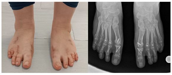
Antibiotics, analgesics, low-molecular-weight heparin, and vitamin D were prescribed for the first 10 days. Serial wound care was performed over 5 weeks, using a non-removable posterior splint. At week five, the fixation pins from the osteotomies and arthrodesis were removed, and by week six, the patient transitioned to wearing rigid-soled athletic shoes and began gait rehabilitation. Six months later, the patient showed complete recovery, with good clinical and radiological progression.
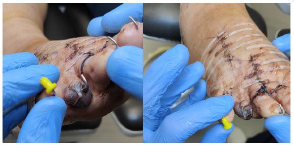
On August 23, 2023, it was decided to operate on the right foot following the same surgical plan. However, during the first dressing change, partial necrosis of the fourth toe was detected due to excessive lengthening of the metatarsal. The fixation pin was removed early, and vasodilators were administered to improve blood circulation. After delimiting the necrosis, partial excision of the fourth toe was performed at eight weeks, removing part of the distal phalanx and the nail unit.
Despite this complication, one year later, the patient shows good clinical recovery and resolution of symptoms.
Discussion
Brachymetatarsia is a congenital deformity characterized by the shortening of one or more metatarsals, affecting both the functionality and appearance of the foot. Although its prevalence varies, it is most commonly seen in the fourth metatarsal. Over the past few decades, new surgical techniques have been developed to treat this condition, ranging from gradual bone distraction to one-stage osteotomy with bone grafting [7,14].
This article analyzes a case of bilateral brachymetatarsia treated with oblique osteotomy using an autologous calcaneal graft, highlighting the complications and outcomes while comparing the various surgical techniques currently available.
One-stage osteotomy is a popular approach for treating brachymetatarsia, allowing for immediate lengthening of the affected metatarsal without the need for multiple surgeries or prolonged periods of gradual bone distraction [9,11,13]. In the case presented, an oblique osteotomy with an autologous calcaneal graft was chosen, a technique that offers several advantages.
According to Sepúlveda et al. (2024), oblique osteotomy is an effective method for correcting deformities ranging from moderate to severe, as it provides a larger bone surface for graft fixation and facilitates faster bone consolidation. In this case, the use of autologous grafts from the calcaneus reduces the risk of rejection and improves graft integration with the recipient bone. This technique is particularly beneficial for patients requiring significant lengthening while avoiding complications associated with allografts [1].
Although combined osteotomy techniques have advantages, one of the main risks is excessive metatarsal lengthening, which can lead to vascular complications. In this case, digital necrosis was observed in one foot, likely caused by excessive lengthening [14-16]. The literature documents this as a potentially serious complication, as exceeding a safe lengthening limit can compromise distal tissue vascularization [15].
Barik et al. (2021) note that maintaining a balance between preserving distal vascularization and achieving adequate functional length is one of the primary challenges of bone-lengthening techniques. Digital necrosis can lead to a loss of functionality and may require additional interventions to preserve tissues [2].
Gradual bone distraction is primarily used for patients requiring more substantial lengthening. The technique described by Machado et al. (2021) involves applying an external fixator after performing an osteotomy to gradually lengthen the affected metatarsal [8]. Although it allows for precise control of length ening and reduces the risk of vascular complications, it has the disadvantage of being a prolonged procedure that requires an extended recovery period.
The use of external fixators can also cause patient discomfort and increase the risk of infections at the pin insertion sites [17,18].
Another surgical option is wedge osteotomy, which involves cutting the metatarsal and inserting a bone graft to lengthen the bone [8,13,14]. According to Fusini et al. (2022), this technique is suitable for moderate lengthenings and allows for effective graft consolidation. However, the risk of pseudoarthrosis or delayed consolidation, especially with allograft use, is one of the main challenges of this technique [5].
One-stage osteotomy without a bone graft is another option. This technique involves performing an osteotomy of the metatarsal and then fixing the bone without grafts using plates or screws [4,5,17]. While less invasive and avoiding the risks associated with grafting, its use is limited to cases requiring minimal lengthening. Cho et al. (2020) indicate that for lengthenings under 10 mm, one-stage osteotomy without grafting can be effective, but for more severe deformities, a graft is necessary to ensure adequate stability [6].
Despite the complication of digital necrosis in one foot, the final outcome in the presented case was satisfactory. After an appropriate follow-up period, the calcaneal graft successfully integrated into both affected metatarsals, and the patient regained both the functionality and appearance of the foot [15,19]. According to the literature, the functional and aesthetic outcomes of one-stage lengthening procedures with grafting are generally favorable when cases are carefully selected, and the limitations of bone lengthening are respected [18-20].
Lamm et al. (2023) propose a classification system for brachymetatarsia based on the severity of the deformity and suggest that one-stage osteotomy with grafting is suitable for moderate to severe cases, provided that lengthening limits are maintained to prevent complications [4].
Conclusion
Oblique osteotomy with autologous calcaneal grafting is an effective technique for treating brachymetatarsia, particularly in cases requiring significant lengthening. Despite the occurrence of digital necrosis in the affected metatarsal toe, the final outcomes were satisfactory. When compared to other techniques such as gradual bone distraction, this method provides a safe and effective option, though it is crucial to avoid excessive lengthening to minimize vascular risks.
References
- Sepulveda M, Orellana G, Sanchez F, Birrer E. Brachymetatarsia. EFORT Open Rev. 2024; 9(4): 235-240. doi: 10.1530/EOR-23-0011.
- Barik S, Farr S. Brachymetacarpia and brachymetatarsia: Do we need to operate? EFORT Open Rev. 2021; 6(1): 15-23. doi: 10.1302/2058-5241.6.200087.
- Córdoba-Fernández A, Vera-Gómez ML. Literature Review on Brachymetatarsia. Orthop Nurs. 2018; 37(5): 292-302. doi: 10.1097/NOR.0000000000000487.
- Lamm BM, Lamm TB. Brachymetatarsia: A Classification for Surgical Treatment. J Foot Ankle Surg. 2023; 62(1): 132-149. doi: 10.1053/j.jfas.2022.06.002.
- Fusini F, Alessandro M, Rava A, Kristijan Z, Felli L, et al. Aetiology, diagnosis, and treatment of brachymetatarsia: a narrative review. Musculoskelet Surg. 2022; 106(2): 99-109. doi: 10.1007/s12306-021-00720-z.
- Cho YJ, Lee JY, Lee WG, Jung ST. Brachymetacarpia and Brachymetatarsia in Patients with Multiple Hereditary Exostosis. Clin Orthop Surg. 2020; 12(4): 549-553. doi: 10.4055/cios19121.
- Caldiño-Lozada I, Gallegos-de la Torre O, Esperón-Hernández R. Tratamiento integral del antepié con doble braquimetatarsia bilateral en un tiempo quirúrgico. Reporte de un caso [Case report: comprehensive treatment of forefoot with double brachymetatarsia in one surgical stage]. Acta Ortop Mex. Spanish. 2017; 31(2): 86-90. PMID: 28840674.
- Machado Filho A, Costa GFO, Faria AADS, Leão GT, Martins JS. Treatment of Brachymetatarsia by Distraction Osteogenesis Using Monolateral External Fixator. Rev Bras Ortop (Sao Paulo). 2021; 57(1): 75-81. doi: 10.1055/s-0041-1735828.
- Toporowski G, Thiesen R, Gosheger G, Roedl R, Frommer A, et al. Callus distraction for brachymetatarsia - A comparison between an internal device and the external fixator. Foot Ankle Surg. 2022; 28(8): 1220-1228. doi: 10.1016/j.fas.2022.04.001.
- Arceri A, Mazzotti A, Zielli SO, Artioli E, Viroli G, et al. What’s the evidence on surgical treatment for congenital brachymetatarsia: A systematic review and meta-analysis. J Orthop. 2023; 49: 107-116. doi: 10.1016/j.jor.2023.11.046.
- Marwan Y, Dalu KA, Bernstein M, Hamdy RC. Metatarsal Gradual Lengthening for Brachymetatarsia Using a Mini-rail External Fixator System. Strategies Trauma Limb Reconstr. 2022; 17(3): 184-188. doi: 10.5005/jp-journals-10080-1565.
- Zhu D, Lefèvre M, Fernandez A, Galois L. Brachymetatarsia: Surgical Management, Case Report, and Literature Review. Case Rep Orthop. 2022; 2022: 8253096. doi: 10.1155/2022/8253096.
- Klauser HO. The Surgical Treatment of Brachymetatarsia. Foot Ankle Clin. 2021; 26(4): 685-704. doi: 10.1016/j.fcl.2021.07.003.
- Guizar-Cuevas S, Mora-Ríos FG, Mejía-Rohenes LC, López-Marmolejo A, Cortés-Gómez J. Elongación por callotaxis en braquimetatarsia congénita [Elongation with callotaxis for congenital brachymetatarsia]. Acta Ortop Mex. Spanish. 2010; 24(6): 395-9. PMID: 21400762.
- Montero-Quijano M, Mora-Ríos FG, Mejía-Rohenes C, López-Marmolejo A, Tamayo-Pacho F, et al. Estudio comparativo de manejo de braquimetatarsia congénita mediante elongación por callotaxis [Comparative study of the management of congenital brachymetatarsia with elongation with callotaxis]. Acta Ortop Mex. Spanish. 2015; 29(2): 77-81. PMID: 27012080.
- Shecaira AP, Fernandes RMP. Brachymetatarsia: One-stage Versus Two-Stage Procedures. Foot Ankle Clin. 2019; 24(4): 677-687. doi: 10.1016/j.fcl.2019.08.010.
- Jagani N, Fagir M, Dalal N, James LA. Metatarsal lengthening by distraction osteogenesis for brachymetatarsia in paediatric patients: is it safe and effective? Eur J Orthop Surg Traumatol. 2024; 34(4): 1839-1844. doi: 10.1007/s00590-024-03860-7.
- Lee KB, Yang HK, Chung JY, Moon ES, Jung ST. How to avoid complications of distraction osteogenesis for first brachymetatarsia. Acta Orthop. 2009; 80(2): 220-5. doi: 10.3109/17453670902930040.
- Robinson JF, Ouzounian TJ. Brachymetatarsia: Congenitally short third and fourth metatarsals treated by distraction lengthening--a case report and literature summary. Foot Ankle Int. 1998; 19(10): 713-8. doi: 10.1177/107110079801901012.
- Fuiano M, Mosca M, Caravelli S, Di Liddo M, Grassi A, et al. Callus distraction with external fixator for the treatment of congenital brachymetatarsia of the fourth ray. Foot Ankle Surg. 2020; 26(6): 693-698. doi: 10.1016/j.fas.2019.08.018.